| |
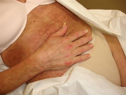
Image 1

Image 2
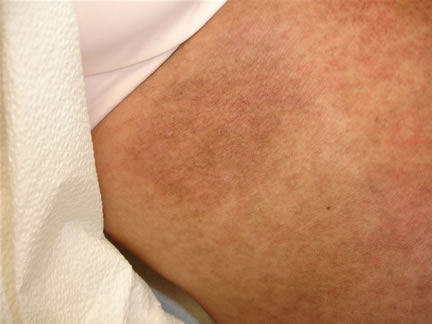
Image 3
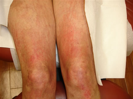
Image 4
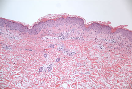
Image 5-Low power magnification shows minimal histopathologic changes.
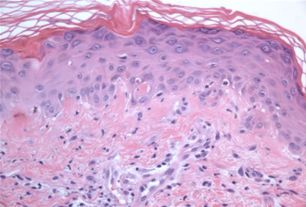
Image 6-Higher power magnification reveals a cell-poor interface dermatitis with vacuolar alteration at the dermal-epidermal junction and dyskeratotic keratinocytes.
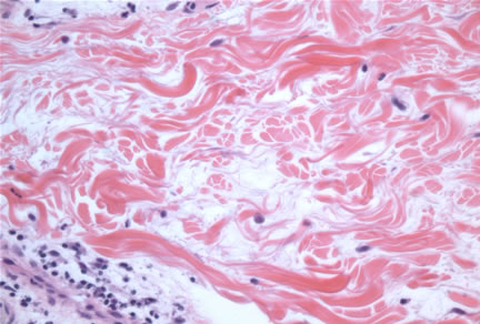
Image 7-The reticular dermis shows separation of the collagen fibers by a diffuse increase in mucin.
What is your diagnosis? |
|
 |
|
Case Study
This is a 52 year old woman who presents with a six month history of arthritis and oral ulcerations. She now presents with a diffuse poikilodermatous rash on her trunk and extremities. (Images 1-4). Her punch biopsy revealed a cell-poor interface dermatitis with dermal mucinosis (Images 5-7).
She is ANA negative and Ro+ by serum serological studies. |