| |
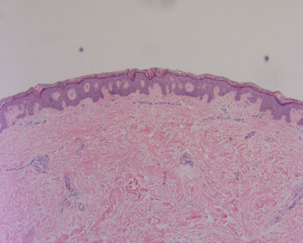
Figure 1-Low power magnification shows mild epidermal hyperplasia with fusion of the rete ridges. The dermis is unremarkable with no significant collagen alterations, lack of an inflammatory infiltrate, and preservation of the adnexal structures.
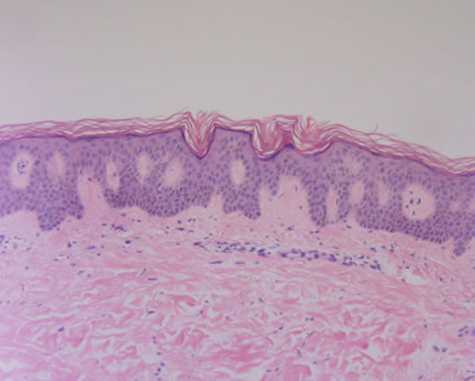
Image 2 -Higher power magnification shows compact orthokeratosis and uniform acanthosis of the epidermis.
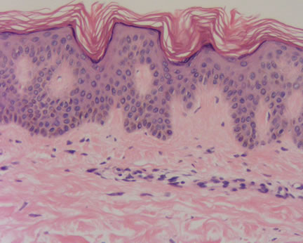
Image 3-The basal epithelial layer shows an increase in melanin pigment unassociated with an increase in melanocytes.
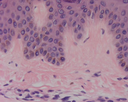
Image 4-Highest power magnification showing the cytologic blandness of the keratinocytes. No interface dermatitis is present. Note the dermis shows a non-specific and minimal perivascular lymphocytic infiltrate with rare melanophages.
What is your diagnosis? |
|
 |
|
Case Study
This is a 27 year old female referred by her primary care provider for evaluation of persistent dryness and discoloration on the right pretibial area present for approximately 2 years. Cortisone cream was prescribed with minimal change. She now presents with an approximately 6 x 10-cm area of reticulate hypo and hyperpigmentation on the right pretibial area with 2 smaller adjacent areas.
The clinical assessment is discoid lupus erythematosus versus morphea.
|