| |
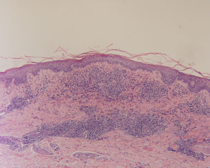
Figure 1-Low power magnification showing a superficial and deep perivascular infiltrate.
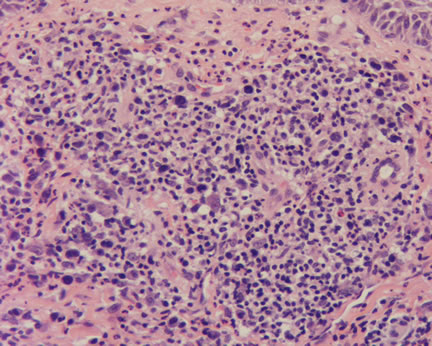
Image 2 -Higher power magnification shows larger, atypical cells admixed with chronic inflammatory cells.
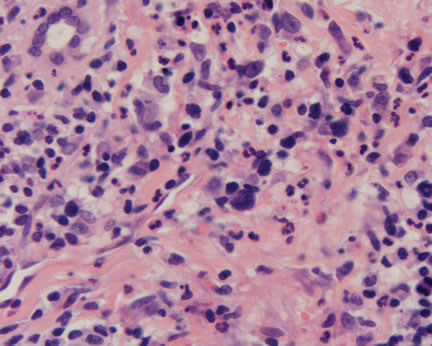
Image 3-The larger atypical cells have prominent nucleoli and occasional angulated nuclei. Note the background inflammatory cell infiltrate consisting of benign lymphocytes, eosinophils, neutrophils, and plasma cells.
What is your diagnosis? |
|
 |
|
Case Study
This is a 42 year old woman with diffuse papules distributed over the trunk and arms.
|