| |
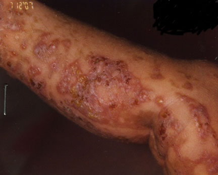
Image 1-Blistering lesions along the lower limb.
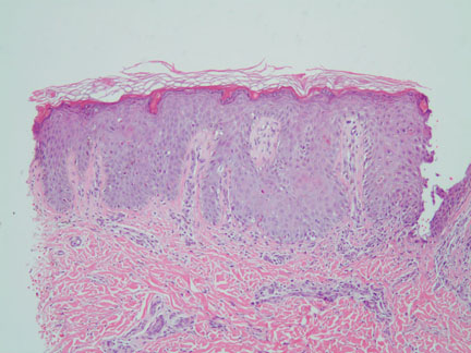
Image 2 -Low power (100x) magnification demonstrates mild epidermal hyperplasia.

Image 3-Higher magnification (200x) demonstrates scattered dyskeratotic keratinocytes.
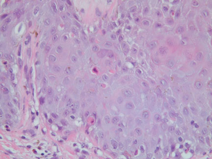
Image 4-Highest magnification (400x) demonstrating the dyskeratotic keratinocytes with minimal accompanying inflammatory cell infiltrate.
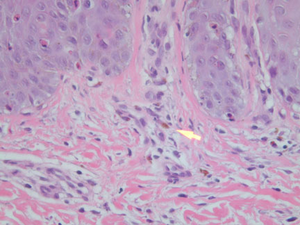
Image 5-Highest magnification demonstrating scattered melanophages.
What is your diagnosis? |
|
 |
|
Case Study
This is a 2 week old girl who presented with unilateral blistering lesions on the extremities. A punch biopsy is taken from the lower leg.
|