| |
Archived Cases |
|
 |
|
Case Study
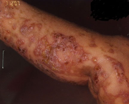
This is a 2 week old girl who presented with unilateral blistering lesions on the extremities.
Diagnosis:
Incontinentia pigmenti-vesicular stage
Discussion
This is a classic example of this rare genodermatoses, inherited as an X-linked dominant gene; most males die in utero. This genetic defect has now been isolated to a mutation in the IKK-gamma gene (NEMO).
Patients may present with a striking sequence of cutaneous lesions beginning with a vesiculobullous lesions, usually in a linear arrangement on the extremities and trunk. The lesions may evolve to a verrucous stage. The third stage presents with striking streaks and whorls of brown to gray hyperpigmentation. A fourth stage may occur many years later with hypochromic lesions, usually on the lower extremities.
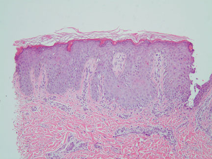
The histopathology mirrors the clinical stages. Eosinophilic spongiosis with intraepithelial vesicles are present in the first stage. If a biopsy is taken between vesicles, numerous dsykeratotic keratinocytes may be present (as in this current case). In the second verrucous stage, there is more pronounced epidermal hyperplasia with papillomatosis and markedly increased dyskeratotic keratinocytes. There is a general decrease in eosinophils with scattered melanophages. In the whorled hyperpigmentation stage, dermal melanophages are abundant.
The striking clinical presentation usually leaves little doubt about the histopathologic diagnosis. However, without the clinical history, and depending upon the clinical stage of the disease, these histopathological findings may also be seen in a variety of diseases that encompass eosinophilic spongiosis (hypersensitivity reactions, urticarial variants of bullous pemphigoid or pemphigus vulgaris, etc), and post-inflammatory pigmentary alterations.
References:
The Doctor's Doctor-Incontinentia Pigmenti |