| |
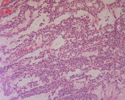 Image 1-Most of the tumor is composed of this cellular and vascular proliferation. Image 1-Most of the tumor is composed of this cellular and vascular proliferation.
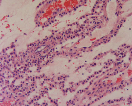
Image 2

Image 3-In one portion of the tumor, a highly cellular nodule is noted, merging with the earlier described areas.
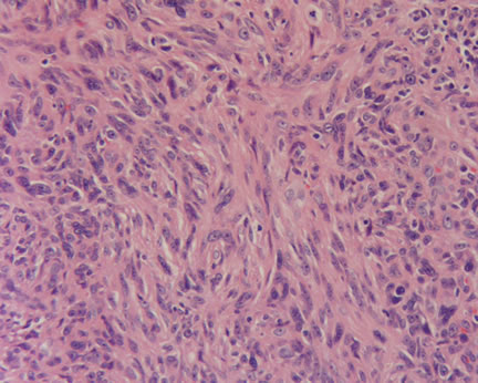 Image 4-Higher power magnification reveals considerable pleomorphism. Image 4-Higher power magnification reveals considerable pleomorphism.
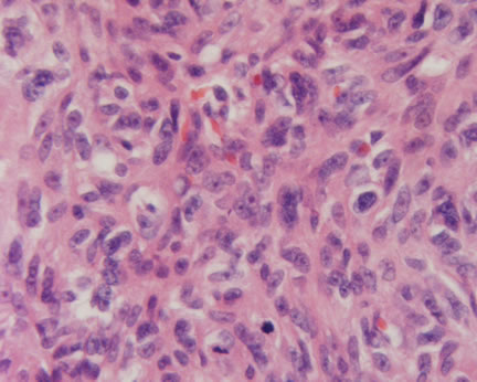 Image 5-At least 5 mitotic figures per 50 high power fields are identified. Image 5-At least 5 mitotic figures per 50 high power fields are identified.
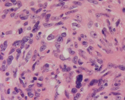
Image 6
What is your diagnosis? |
|
 |
|
Case Study
This is a 65 year old man who presents with a 3 cm nodule on the left elbow.
|