| |
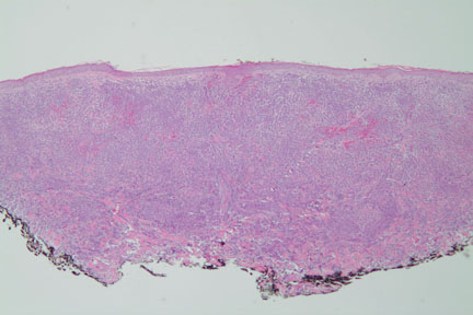
Image 1-Low power magnification shows a diffuse dermal infiltrate.
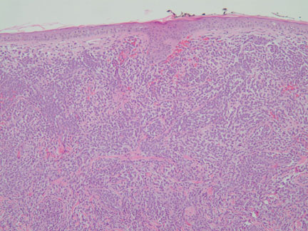
Image 2-The infiltrate is separated from the epidermis by a Grenz zone.
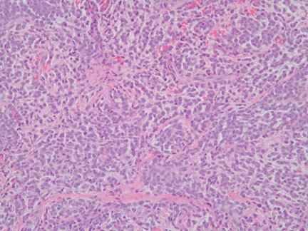
Image 3-Mononuclear cells are interspersed with extravasated red blood cells.
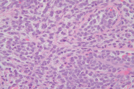
Image 4-The mononuclear cells are small and mitotically active.
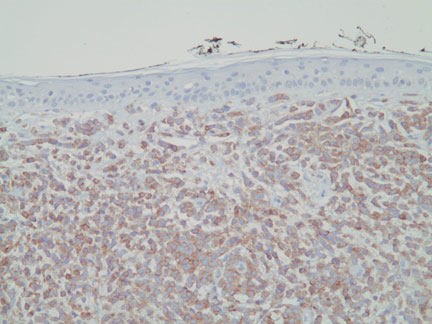
Image 5-All of the cells are diffusely positive for CD45 (Leukocyte common antigen) and negative for cytokeratin and S100.
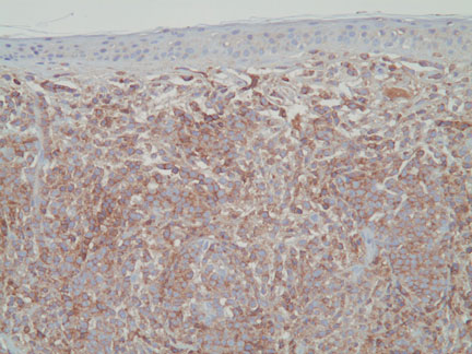
Image 6-Diffuse positivity for CD56 (natural killer cell phenotype)
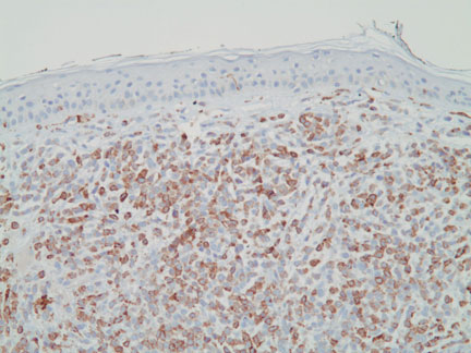
Image 7-Diffuse positivity for CD68.
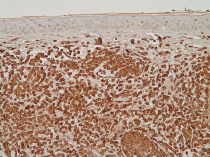
Image 8-Diffuse positivity for myeloperoxidase (MPO).
The tumor cells were negative for CD3, CD20, CD33, CD34, CD79a, CD99, CD117, and TdT.
What is your diagnosis? |
|
 |
|
Case Study
This is a 85 year old woman who presents with multiple ecchymotic plaques on the upper trunk.
|