Case Study
This is a 85 year old woman who presents with multiple ecchymotic plaques on the upper trunk.
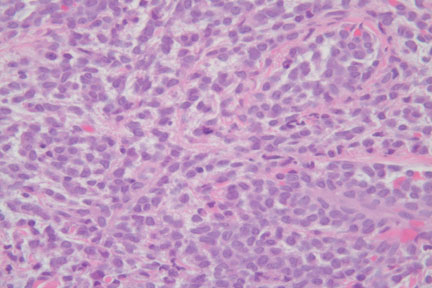
DIAGNOSIS:
Myeloid Sarcoma (Leukemia Cutis)
Comment:
This is a challenging diagnosis. Except for the ecchymoses, the patient experienced no constitutional signs of her underlying disease. The histopathology may be mimicked by several neoplastic processes including a peripheral T-cell lymphoma, a small cell carcinoma, a small cell melanoma, and a wide variety of tumors collectively known as small round blue cell tumors. Although these latter tumors are more common in childhood (such as neuroblastomas, primitive neuroectodermal tumors/Ewing's sarcoma, and rhabdomyosarcomas), in selected cases, these differential diagnostic considerations may enter into the differential diagnosis of adult tumors.
Leukemia cutis is the cutaneous involvement by neoplastic leukocytes. Since the latter may be either myeloid or lymphoid, and either an acute or chronic presentation, a wide variety of subtypes may secondarily involve the skin. Historically, the term granulocytic sarcoma or chloroma describes leukemic infiltrates derived from granulocytic precursors whereas monocytic precursors may be termed monoblastic sarcoma. Myeloid sarcoma and extramedullary myeloid tumor refers to tumors derived from both granulocytic and monocytic precursors.
The histopathologic pitfalls are rendered even more challenging by the varied immunohistochemical staining patterns. The following table adapted from Pileri, etal, highlights the frequency that one may expect to find these immunostains:
| Stain |
Percentage |
| CD68 |
100% |
| Myeloperoxidase |
83.6% |
| CD117 |
80.4% |
| CD99 |
54.3% |
| CD34 |
43.4% |
| TdT |
31.5% |
| CD56 |
13% |
| CD30 |
2.2% |
| CD4 |
1.1% |
FISH analysis for chromosomal aberrations may also aid in the diagnostic workup with slightly greater than half of the cases exhibiting a common leukemic aberration.
The dermatologist should also be aware that leukemia cutis may rarely present without peripheral blood or bone marrow involvement by the leukemia, so-called aleukemic leukemia cutis. In these cases, the skin or mucosal lesions may precede the bone marrow or peripheral blood involvement by several weeks to months, necessitating careful clinical evaluation and follow-up.
Submitted by Paul K. Shitabata, M.D.
Director of Dermatopathology
Harbor-UCLA Dermatology
References:
Leukemia Cutis. Am J Clin Pathol 2008;129:130-142. Cho-Vega JH, etal.
Myeloid sarcoma: clinico-pathologic, phenotypic and cytogenetic analysis of 92 adult patients. Leukemia. 2007 Feb;21(2):340-50. Pileri SA, etal.
The Doctor's Doctor-Leukemia
|