| |
Melanoma

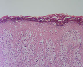
Melanocytic Nevus
|
|
 |
|
Presentations
Congenital Melanocytic Nevus-Clinical and Pathologic Pitfalls
Dysplastic Nevus-Atypical Mole or Typical Role?
Is this Melanoma? Should it Be?
Melanoma-Back to Basics, I Thought I Knew Ya!
Melanoma in Situ-Taking it to the Lowest Level
Melanoma Sentinel Lymph Node Biopsy-Is it the Gold Standard?
Melanoma Through the Malpractice Scope
The Spitz Nevus-Sophie Spitz Was Correct!
Subungual Melanoma
This is a Melanoma? Unusual Histologic Variants from the Great Mimic!
Web Links
Argyria
Becker's Nevus (Becker's Hairy Nevus)
Blue Nevus
Congenital Nevus
Dysplastic Nevus
Hypopigmented Lesions of the Skin including Vitiligo
Idiopathic Eruptive Macular Pigmentation (IEMP)
Lentigo
Melanocytic Matricoma
Melanoma
Melanoma, Non-Skin Tumors
Melanoma Histopathological Variants/Special Stains/Differential Diagnosis
Melanoma Prognostic Factors
Melanoma Treatment
Mole (Pigmented Nevus)
Nevus of Ota and Ito
Pigmented Spindle Cell Nevus of Reed
Reticular Melanotic Hypermelanosis
Spitz Nevus
Tattoo
Journal Articles
Three Dimensional Visualization of Lymphatic Drainage Patterns in Patients with Cutaneous Melanona. Lancet Oncol 2007;8:806-812. Reynolds HM, etal.
Reliability of Lymphatic Mapping After Wide Local Excision of Cutaneous Melanoma. Ann Surg Oncol 2007;12:2377-2383. Ariyan S, etal.
Are En Face Frozen Sections Accurate for Diagnosing Margin Status in Melanocytic Lesions? Am J Clin Pathol 2003;120:203-208. Prieto VG, etal.
|