|
|
|
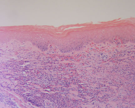
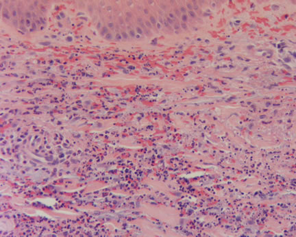
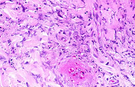
Issues in Dermatopathology A report of 4 patients with late-onset LV probably due to warfarin It is a pleasure to read an article written by a current resident. In this article, Dr. Binh Ngo, one of our current dermatology resident, brings to our attention the unusual clinical manifestation of a late-onset leukocytoclastic vasculitis secondary to warfarin therapy. An interesting and perhaps controversial point of the paper is the long time that elapsed between the initiation of the warfarin and the onset of the vasculitis, ranging from 2-10 years. Significantly, in 2 of the patients, there was the onset of the lesions following a rechallenge. The lesions recurred between 5 and 23 days after rechallenge, supporting the authors' hypothesis. Given the widespread use of warfarin, it is somewhat surprising that a review of the literature reveals only three other cases. One suspects this condition may be under-reported or under-recognized. It certainly deserves greater attention and we thank Dr. Ngo and her colleagues for their careful and provocative paper. When you have reviewed the case and discussion, please comment, I will post them anonymously. Additional References: The Doctor's Doctor-Leukocytoclastic Vasculitis
|
Last Updated August 2, 2005
Send Emails to
Webmaster at DermpathMD
Read the Medical Disclaimer