|
|
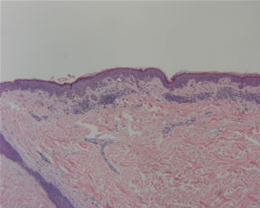
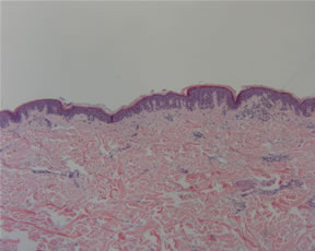
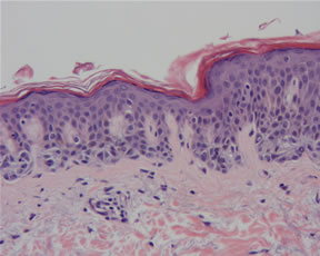
The Atypical Melanocytic Nevus AKA The Dysplastic Nevus You may be astounded over the names that the atypical nevus has been called. Dysplastic nevus And despite our best attempts to give it an identity, there is still tremendous controversy over its very terminology. What is dysplasia? The dermatology, dermatopathology, and pathology literature are replete with articles and editorial postings seeking to define and quantify dysplasia. This article will not add to these erudite discussions but rather will discuss one approach. We prefer the term atypical melanocytic nevus and qualify it with the degree of atypia, usually a low-grade or high-grade atypia. Atypia is non-committal and is a descriptive diagnosis that alerts the treating physician that this is a lesion that may require careful follow-up as well as consideration for a complete removal. It also takes into account the research that has examined the specificity of both clinical and histopathologic features that have classically been associated with the dysplastic nevus. One study by Dr. Barr and colleagues examined fifty-eight junctional and compound nevi from 26 subjects. All nevi met the clinical criteria of benign common nevi: 5 mm or less in diameter, symmetric, round or slightly oval, uniform pigmentation, distinct and regular margins, and no erythema. Microscopic examination revealed one or more of the histologic features associated with dysplastic nevi (disordered bridging nests, papillary dermal fibrosis with inflammation, melanocytic atypia) with the following frequencies: At least one: 87.8% Clearly, the histologic features of dysplastic nevi commonly occur in benign common acquired nevi. The converse of this study was examined by Dr. Urso who examined six histologic features (dimension > 5 mm, lentiginous proliferation, disordered nested pattern, melanocytic dyskaryosis, dermal lymphocytic infiltrate, suprabasal melanocytes) in 253 melanocytic nevi with different clinical appearances. Atypical histologic features, found in 72% of nevi, occurred singly or formed numerous and highly variable combinations. Dr. Urso found a complex histologic spectrum comprising lesions showing a progressively increasing incidence of atypical features rather than two classes (common and dysplastic nevi). In fact, he identified a total of 5 distinct histopathologic classes and concluded that diagnostic categories such as dysplastic nevi and common nevi are inappropriate because they do not adequately reflect the complexity of these lesions. For my tounge in cheek contribution, I have mentioned to my colleagues that we should discard all of the above terminology and utilize the system of MUMP...melanocytic nevus of uncertain malignant potential. For pathologists, there is obvious analogy with tumors in other organ system such as gastrointestinal stromal tumors or smooth muscle tumors of the uterine corpus. However, for many, the groans that are already being emitted will no doubt drown out my suggestion and lead to a premature and some say, well-deserved death. 'Tis a pity, it is a much catchier phrase than the BK Mole! Submitted by Paul K. Shitabata, M.D. References: Urso C. Atypical histologic features in melanocytic nevi. Am J Dermatopathol 2000; Oct;22(5):391-6. Klein LJ and Barr RJ. Histologic atypia in clinically benign nevi. A prospective study. J Am Acad Dermatol 1990 Feb;22(2 Pt 1):275-82 |
Last Updated
March 10, 2006
Send Emails to
Webmaster at DermpathMD
Read the Medical Disclaimer