|
|
|
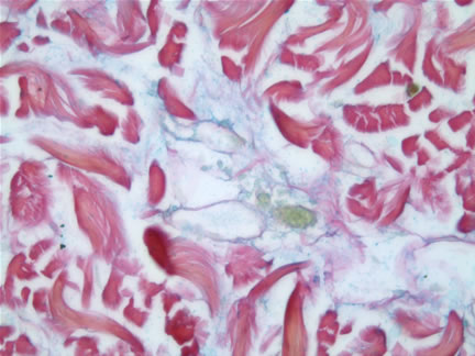
Case Study: Reticular Erythematous Mucinosis This case brings together a variety of interesting clinical and histopathological features. The name of the disease perfectly describes both features. There is nothing specific about a reticulated erythematous rash. It may be a primary skin disease or part of a systemic disorder. Classic diagnoses include: Confluent and reticulated papillomatosis Similarly, the histopathologic findings of a superficial and deep perivascular and periadnexal lymphocytic infiltrate with dermal mucinosis may be visualized with several dermatological disorders including: Lupus Since the common link in these two categories is lupus erythematosus, it was critical to exclude this latter disease. In this current case, the patient's ANA was negative. A biopsy for direct immunofluorescence was not performed. It should be noted that some investigators do believe REM to be a variant of lupus. This case illustrates the cooperative marriage between the clinical and histopathologic findings, to arrive at a diagnosis. Now, if only we could just transfer this harmony to humans! Clinical photo and case history submitted by Elizabeth Lener, M.D., Ladera Ranch, CA. References: The Doctor's Doctor-Reticular Erythematous Mucinosis Comments: |
First Posted March 30, 2006
Archived Case Studies
Send Emails to
Webmaster at DermpathMD
Read the Medical Disclaimer