| |
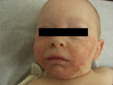
Figure 1
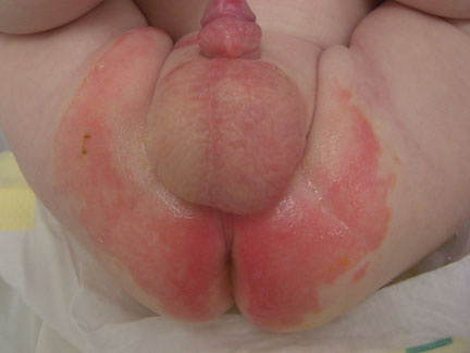
Figure 2
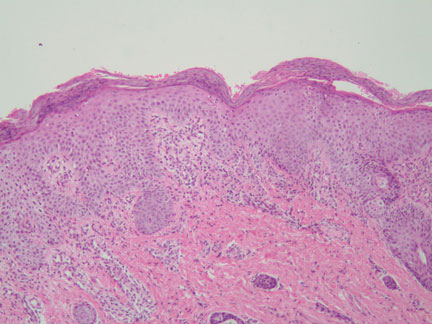
Figure 3-Low power photomicrograph taken from skin lesions (100x).

Figure 4-Medium power magnification (200x).
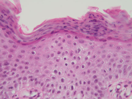
Figure 5-Highest magnification (400x)
What is your diagnosis? |
|
 |
|
Case Study
This is a 4 month old boy, born at 30 weeks gestation due to premature rupture of membranes. He presents with a 2 month history of well-demarcated, erosive, vivid red plaques on face and buttocks. The mother described the appearance of "blisters". He was very irritable, crying throughout the entire visit.
His medical history is significant for secondary Candida and Staph infection, treated with antifungals, topical steroids and oral antibiotics.
|