| |
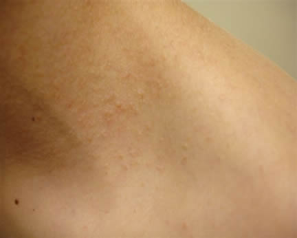
Image 1-Slightly raised macular-papular eruption on the neck.
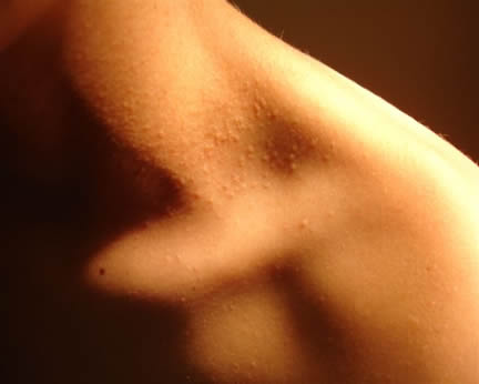
Image 2 -Contrast photograph
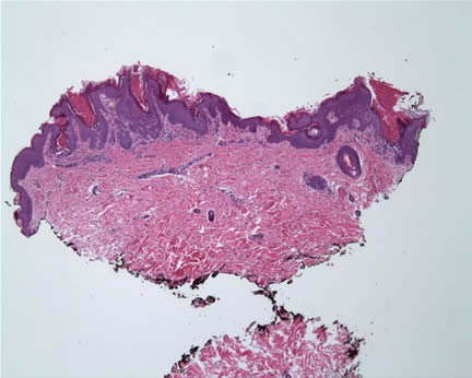
Image 3-Low power magnification showed variable epidermal hyperplasia.
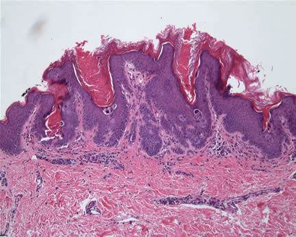
Image 4-Higher magnification reveals follicular plugging associated with acantholysis.
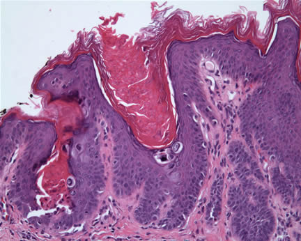
Image 5-Acantholytic cells are predominately distributed within the stratum corneum, subcorneum, and stratum granulosa.
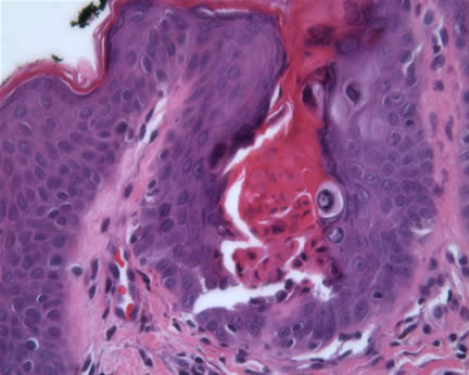
Image 6-Corp ronds and corp grains are noted.
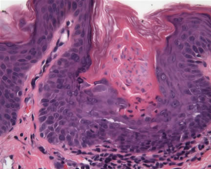
Image 7-In rare foci, cornoid lamellae are noted.
What is your diagnosis? |
|
 |
|
Case Study
This is a 10 year old boy who was treated with topical steroids for presumed eczema. The lesions were asymptomatic but worsened with the topical treatment. The lesions were limited to the neck. The patient is in otherwise good health.
|