| |
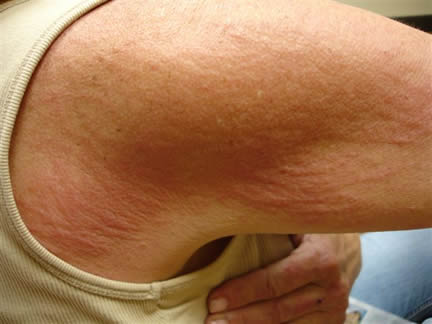
Image 1
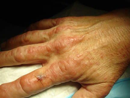
Image 2
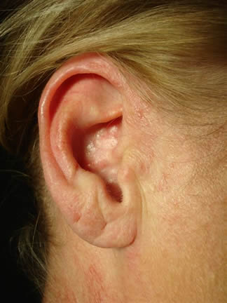
Image 3
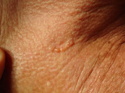
Image 4
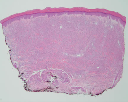 Image 5-Low power magnification showing diffuse replacement of the dermis by a cellular infiltrate, separated from the epidermis by a Grenz zone. Image 5-Low power magnification showing diffuse replacement of the dermis by a cellular infiltrate, separated from the epidermis by a Grenz zone.
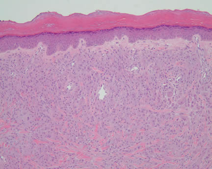 Image 2 Image 2
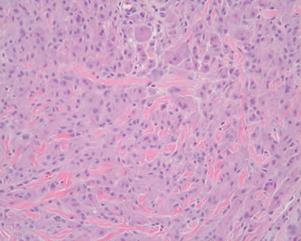 Image 3-Higher power magnificatioin exhibiting histiocytic appearing cells with occasional giant cells. Image 3-Higher power magnificatioin exhibiting histiocytic appearing cells with occasional giant cells.
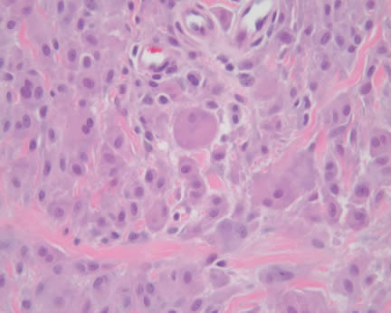 Image 4 Image 4
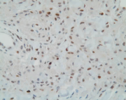
Image 5-Diffuse nuclear positivity for p53.
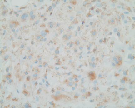 Image 6-Diffuse immunopositivity for CD68 Image 6-Diffuse immunopositivity for CD68
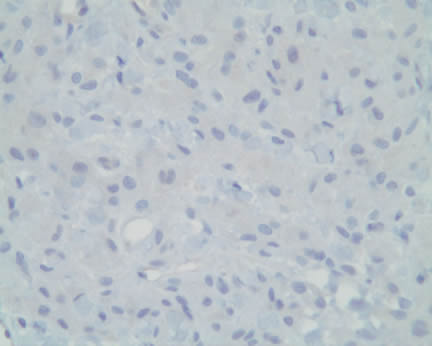
Image 7-Immunohistochemical stains for CD1a, Cytokeratin, S100, and smooth muscle actin were negative. CD45 revealed focal positivity.
What is your diagnosis? |
|
 |
|
Case Study
This is a 56 year old woman who presents with an acute onset of multiple erythematous papules occurring on the proximal extremities, trunk, head, and neck.
|