| |
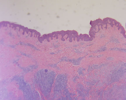 Image 1-Most of the tumor is centered within the deeper dermis with minimal epidermal alterations. Image 1-Most of the tumor is centered within the deeper dermis with minimal epidermal alterations.
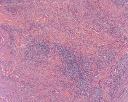
Image 2 -The infiltrate has varying areas of cellularity with areas of sclerosis alternating with denser cellular nodules.
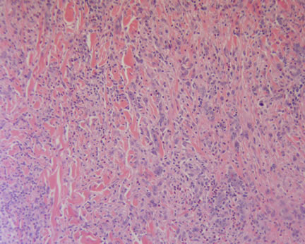
Image 3-The cytology of the infiltrate varies from larger epithelioid appearing cells with a mixed chronic inflammation.
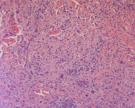 Image 4-Considerable pleomorphism is noted. Image 4-Considerable pleomorphism is noted.
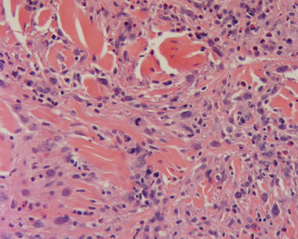 Image 5-Occasional mitotic figures, including atypical mitotic figures are noted. Image 5-Occasional mitotic figures, including atypical mitotic figures are noted.
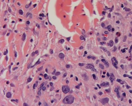
Image 6-Large epithelioid appearing cells with prominent nucleoli are noted admixed with chronic inflammatory cells including plasma cells and eosinophils.
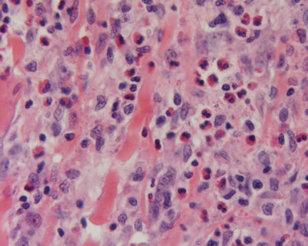
Image 7
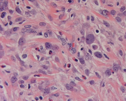 Image 8 Image 8
What is your diagnosis? |
|
 |
|
Case Study
This is a 44 year old man who presents with a tumor in the right hip.
|